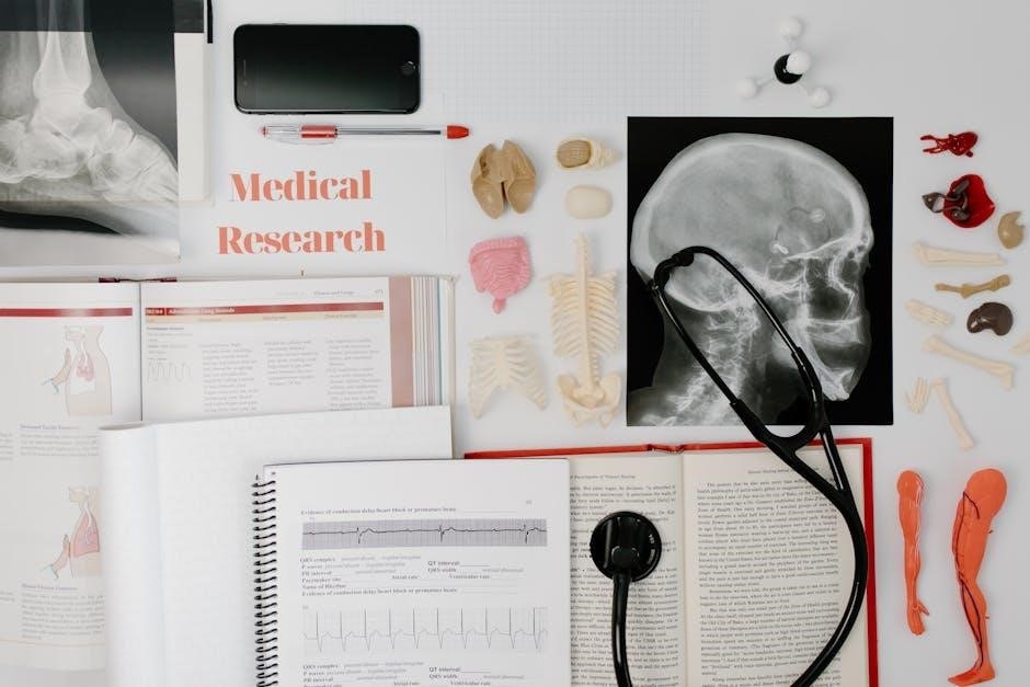
This manual expertly blends human anatomy study with practical cat dissections, offering a comprehensive lab experience. It’s designed for one-semester courses, providing 30 detailed exercises.
Full-color illustrations and a clear writing style aid learning, while tables and clinical connections enhance understanding of mammalian anatomy and physiological systems.
Purpose of the Manual
This laboratory manual serves as a dedicated companion for students undertaking a one-semester human anatomy course, specifically incorporating cat dissections to solidify learning. Its primary purpose is to provide a hands-on, visual, and engaging experience that complements traditional lecture material.
The manual aims to bridge the gap between theoretical knowledge and practical application, allowing students to directly observe and identify anatomical structures in a mammalian model. Through step-by-step dissection instructions and detailed illustrations, it fosters a deeper comprehension of human anatomy by highlighting similarities and differences.
Furthermore, it equips students with essential dissection skills, anatomical terminology, and the ability to relate lab activities to real-world clinical scenarios, enhancing their overall understanding of physiological systems.
Historical Context of Cat Dissections in Anatomy Education
Cat dissections have a long-standing tradition in anatomy education, dating back centuries when human cadavers were difficult to obtain or ethically restricted. Felines, possessing anatomical similarities to humans, provided a readily available and ethically more acceptable alternative for studying mammalian structure.
Historically, cat dissections allowed students to visualize organ systems, muscle attachments, and nerve pathways, building a foundational understanding of human anatomy. This practice continued to evolve alongside advancements in anatomical knowledge and pedagogical techniques.
Even with increased access to human specimens, cat dissections remain valuable for reinforcing concepts, developing dissection skills, and providing a comparative perspective on mammalian anatomy, as evidenced by their continued inclusion in modern lab manuals.
Safety Precautions in the Anatomy Lab
Safety is paramount during cat dissections. Always wear appropriate personal protective equipment (PPE), including safety glasses, gloves, and a lab coat, to prevent exposure to biological materials and potential hazards. Proper handling of dissection instruments – scalpels, scissors, and probes – is crucial to avoid self-injury or damage to specimens.
Maintain a clean and organized workspace, disinfecting surfaces before and after use. Dispose of biological waste properly, following established lab protocols. Be mindful of potential allergens and avoid touching your face during the dissection process.
Report any accidents or spills to the instructor immediately. A thorough understanding and adherence to these safety guidelines ensures a productive and secure lab experience.

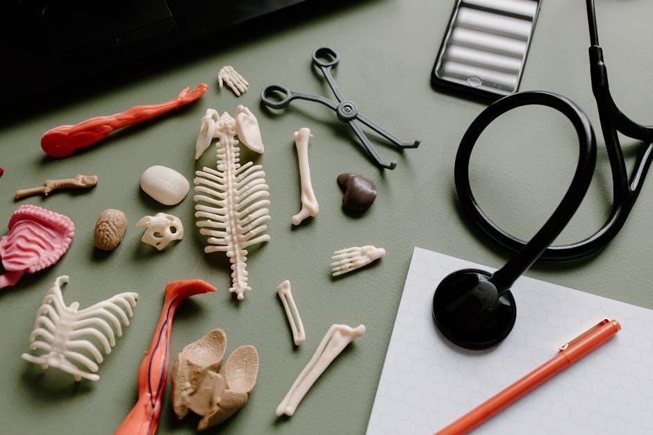
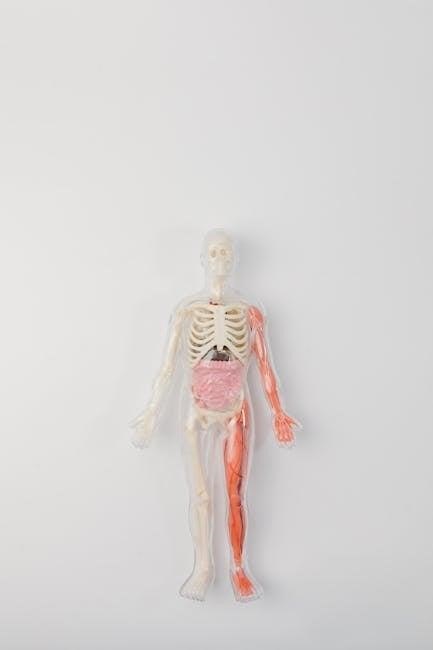
Skeletal System Dissection
Dissection of the cat skeleton reveals striking similarities to human bone structure, aiding comparative anatomy study. Identify major bones and explore joint articulations.
Feline vs. Human Skeletal Similarities
The feline skeleton provides an excellent model for understanding human skeletal anatomy due to fundamental homologies established through evolutionary history. Both species share a similar bone count and arrangement, exhibiting analogous structures in the limbs, vertebral column, and skull.
Comparative osteology highlights shared features like the presence of long bones in the limbs – humerus, radius, ulna, femur, tibia, and fibula – facilitating similar ranges of motion. The vertebral column, composed of cervical, thoracic, lumbar, sacral, and caudal vertebrae, demonstrates conserved segmentation.
However, key differences exist reflecting adaptations to locomotion; cats possess a more flexible spine and a prominent clavicle, while humans have a broader pelvis and a more robust femur for bipedalism. Despite these variations, the cat skeleton effectively illustrates core human anatomical principles.
Identifying Major Bones of the Cat Skeleton
Dissection reveals the cat’s axial skeleton comprising the skull, vertebral column, and rib cage. The skull showcases cranial and facial bones, including the mandible, maxilla, and zygomatic arch. The vertebral column, segmented into cervical, thoracic, lumbar, sacral, and caudal regions, supports the body and protects the spinal cord.
Appendicular skeleton elements include the scapula, humerus, radius, ulna, carpals, metacarpals, and phalanges in the forelimb. Hindlimb bones consist of the pelvis, femur, tibia, fibula, tarsals, metatarsals, and phalanges. Careful observation identifies bone markings like processes, tubercles, and foramina, serving as muscle attachment sites.
Accurate identification of these bones is crucial for understanding feline anatomy and establishing parallels with the human skeletal system, reinforcing comparative anatomical knowledge.
Comparative Osteology: Human Bone Equivalents
Cat skeletal anatomy provides a valuable framework for understanding human osteology. The feline scapula corresponds to the human shoulder blade, while the humerus, radius, and ulna align with the upper arm and forearm bones. Similarly, the pelvis and femur mirror the human hip and thigh bones.
Comparative analysis reveals the cat’s carpals and metacarpals are analogous to the human wrist and hand bones. The vertebral column’s segments – cervical, thoracic, lumbar, sacral, and caudal – have direct human counterparts. Recognizing these homologies enhances comprehension of skeletal structure.
Dissection facilitates identifying these equivalents, solidifying knowledge of both feline and human anatomy, and highlighting evolutionary relationships.
Joints and Articulations in the Cat Skeleton
Cat skeletal joints, like those in humans, enable movement. Dissection reveals various articulation types: fibrous, cartilaginous, and synovial. Synovial joints – ball-and-socket (shoulder, hip), hinge (elbow, knee), and pivot – are prominent, allowing a wide range of motion.
Ligaments stabilize these joints, connecting bone to bone, while tendons attach muscle to bone, facilitating movement. Observing these structures in the cat skeleton clarifies their function in both feline and human anatomy.
Comparative study highlights similarities in joint structure and function, reinforcing understanding of biomechanics and musculoskeletal systems. The manual aids in identifying these key components.
Muscular System Dissection
Dissection reveals cat muscles mirroring human counterparts, aiding anatomical understanding. This section focuses on identifying superficial and deep muscles, their functions, and surrounding fascia.
Superficial Muscles of the Cat: Identification and Function
Carefully expose the superficial muscles, beginning with the skin and subcutaneous tissues of the cat. Identify key muscles like the platysma, a thin sheet covering the neck, and the omohyoid, supporting the hyoid bone. Observe the sternocleidomastoid, crucial for head movement, noting its origin and insertion points.
The pectoralis muscles, responsible for limb movement, are prominent in the chest. Further dissection reveals the deltoid, enabling shoulder flexion, and the biceps brachii and triceps brachii, controlling elbow flexion and extension respectively.
Relate these cat muscles to their human equivalents, understanding shared functions and anatomical similarities. Document observations, noting muscle size, shape, and fiber direction. This reinforces knowledge of human skeletal muscle anatomy through comparative study.
Deep Muscles of the Cat: Identification and Function
Proceeding deeper, carefully dissect to reveal the muscles underlying the superficial layer. Identify the splenius and longissimus muscles, vital for vertebral extension and rotation. Locate the iliocostalis, contributing to lateral flexion of the spine. These muscles form part of the erector spinae group.
Examine the serratus ventralis, aiding in rib elevation, and the transversus abdominis, crucial for core stability. Observe the deeper limb muscles, including the supraspinatus and infraspinatus, involved in shoulder rotation.
Compare these deep cat muscles to their human counterparts, noting functional parallels. Document muscle attachments and actions, enhancing understanding of mammalian musculoskeletal systems. This dissection reinforces anatomical knowledge and comparative anatomy principles.
Muscle Tissue Types: Comparison in Cat and Human
Observe the three muscle tissue types – skeletal, smooth, and cardiac – in the cat. Skeletal muscle, attached to bones, exhibits striations and voluntary control, mirroring human skeletal muscle. Smooth muscle, found in organ walls, lacks striations and operates involuntarily in both species.
Cardiac muscle, exclusive to the heart, displays striations and involuntary rhythmicity, functioning similarly in cats and humans. Examine histological slides to compare fiber arrangements and cellular structures.
Note the similarities in muscle fiber composition and contractile mechanisms. Despite species differences, the fundamental principles of muscle contraction remain consistent, highlighting evolutionary conservation. This comparison reinforces understanding of muscle physiology.
Fascia and Muscle Compartmentalization
Observe how fascia, a dense connective tissue, envelops and separates cat muscles. Superficial fascia underlies the skin, while deep fascia organizes muscles into functional compartments. These compartments contain specific muscle groups, nerves, and blood vessels.
Identify the boundaries of muscle compartments and note how fascia provides structural support and facilitates coordinated movement. Dissection reveals that fascia protects muscles and allows for efficient force transmission.
Compare this compartmentalization to human muscle organization, recognizing the analogous arrangement. Understanding fascia’s role is crucial for comprehending muscle function and clinical implications related to injury or inflammation.
Nervous System Dissection
Explore the cat’s brain, spinal cord, and peripheral nerves, comparing structures to human neurology. Dissection reveals sensory organs – eyes, ears, and nose – and their neural connections.
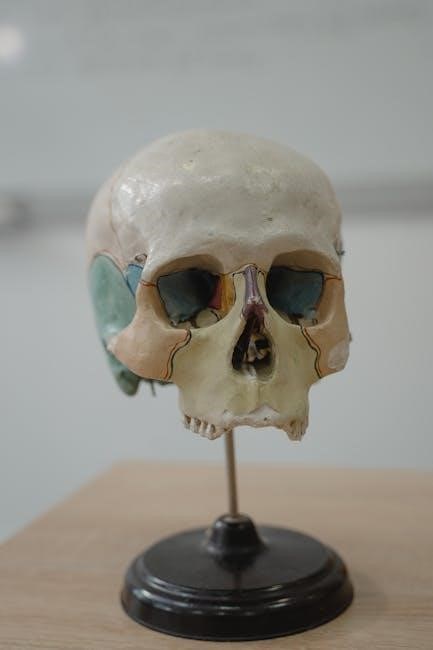
Brain Anatomy: Cat vs. Human
Comparative brain dissection reveals striking similarities and key differences between the feline and human brain. While the basic organization – cerebrum, cerebellum, and brainstem – is conserved, proportional sizes of regions vary significantly.
Observe the relatively smaller cerebral hemispheres in the cat, reflecting a lesser degree of cortical folding (gyrification) compared to humans. Identify homologous structures like the olfactory bulbs, optic lobes, and temporal lobes, noting differences in prominence.
Carefully dissect to expose the midbrain, pons, and medulla oblongata, recognizing their roles in vital functions. This dissection emphasizes the evolutionary adaptations in brain structure correlating with behavioral and cognitive complexity.
Understanding these distinctions provides valuable insight into mammalian neuroanatomy and the basis for higher-order processing.
Spinal Cord and Peripheral Nerves
Dissection reveals the protected location of the spinal cord within the vertebral column. Carefully expose the spinal cord and observe its segmented structure, noting the dorsal and ventral roots emerging from each segment. These roots give rise to the peripheral nerves.
Identify major nerve plexuses – cervical, brachial, lumbar, and sacral – and trace the paths of key peripheral nerves supplying limbs and body wall muscles. Compare the relative size and distribution of nerves in the cat to their human counterparts.
Observe the differences in nerve branching patterns and innervation territories. This dissection highlights the fundamental organization of the central and peripheral nervous systems, crucial for motor control and sensory perception.
Understanding these structures is vital for comprehending neurological function and dysfunction.
Sensory Organs: Eye, Ear, and Nose
Dissection allows for detailed examination of the cat’s sensory organs, revealing adaptations for their predatory lifestyle; Carefully dissect the eye, identifying the cornea, lens, retina, and optic nerve. Compare the tapetum lucidum – a reflective layer – to the human eye’s lack of this feature.
Expose the ear, noting the external ear structures, middle ear ossicles (malleus, incus, stapes), and inner ear cochlea. Observe the nasal cavity, identifying the olfactory epithelium responsible for the sense of smell.
Relate the anatomical structures to their respective functions in vision, hearing, and olfaction. Consider how these sensory systems differ between cats and humans, reflecting their distinct ecological niches.
Understanding these differences enhances appreciation for sensory biology.
Digestive System Dissection
Dissection reveals the cat’s digestive tract – oral cavity to anus – mirroring human systems. Identify the stomach, intestines, liver, pancreas, and gallbladder for comparative study.
Oral Cavity and Esophagus
Begin the digestive system dissection by carefully examining the cat’s oral cavity. Observe the teeth, tongue, and hard/soft palates, noting structural similarities and differences compared to human anatomy. Identify the salivary glands and their ducts, understanding their role in initial food processing.
Next, trace the path of the esophagus, a muscular tube connecting the pharynx to the stomach. Carefully dissect along the esophagus, noting its layers and peristaltic movements. Compare the esophageal structure in the cat to its human counterpart, focusing on muscular composition and mucosal lining. Understand how these structures facilitate the transport of food to the stomach.
Pay attention to the esophageal opening into the stomach, noting its sphincter mechanism.
Stomach and Intestines
Dissect the cat’s stomach, identifying its regions: cardia, fundus, body, and pylorus. Observe the rugae, muscular folds allowing for expansion. Examine the stomach lining, noting the gastric pits and glands responsible for acid and enzyme secretion. Compare the stomach’s structure to that of humans, focusing on functional similarities.
Trace the small intestine – duodenum, jejunum, and ileum – noting its length and coiled structure. Identify the villi and microvilli, enhancing nutrient absorption. Continue to the large intestine – cecum, colon, and rectum – observing its wider diameter and haustra. Relate these structures to their roles in digestion and waste processing, drawing parallels to human anatomy.
Note the differences in intestinal length between cats and humans.
Accessory Digestive Organs: Liver, Pancreas, and Gallbladder
Locate the cat’s liver, the largest internal organ, and identify its lobes. Observe its color and texture, noting its role in bile production, detoxification, and metabolism. Examine the gallbladder, a small sac storing bile, and trace the bile duct connecting it to the duodenum.
Find the pancreas, a glandular organ with both endocrine and exocrine functions. Identify its lobes and relate its position to the duodenum, where pancreatic enzymes aid digestion. Compare these organs to their human counterparts, highlighting structural and functional similarities.
Consider how these accessory organs contribute to overall digestive efficiency in both species.

Respiratory System Dissection
Dissection reveals the cat’s nasal cavity, pharynx, larynx, trachea, lungs, and diaphragm. Compare these structures to human anatomy, noting similarities in respiratory function and pathways.
Nasal Cavity and Pharynx
Begin by carefully exposing the cat’s nasal cavity, observing its internal structures like the nasal septum and conchae, which increase surface area for warming and humidifying inhaled air. Note the connection between the nasal cavity and the pharynx – a shared pathway for air and food.
Compare the feline pharynx to its human counterpart, identifying the nasopharynx, oropharynx, and laryngopharynx. Observe the epiglottis, a crucial structure preventing food from entering the trachea during swallowing.
Consider how these structures contribute to respiration and olfaction in the cat, and relate these observations to the analogous functions in human anatomy. Dissection provides a tangible understanding of these vital respiratory components.
Larynx and Trachea
Carefully dissect to reveal the cat’s larynx, identifying key structures like the thyroid cartilage and vocal folds. Observe how these folds vibrate to produce sound, a function shared with humans, though differing in complexity.
Trace the path of the trachea, noting its cartilaginous rings which provide structural support preventing collapse during breathing. Compare the tracheal structure to its human equivalent, recognizing the similar function of maintaining an open airway.
Examine the branching points where the trachea divides into the bronchi, initiating the respiratory tree. This dissection reinforces understanding of the respiratory pathway and its anatomical components in mammals.
Lungs and Diaphragm
Carefully expose the cat’s lungs, observing their lobed structure and spongy texture. Note the differences in lobe number compared to the human lung, highlighting species-specific adaptations for respiration.
Trace the bronchi as they branch into smaller and smaller airways within the lungs, ultimately leading to the alveoli – the sites of gas exchange. Consider the vast surface area created by these alveoli, crucial for efficient oxygen uptake.
Identify the diaphragm, the muscular sheet separating the thoracic and abdominal cavities. Understand its role in breathing – contraction expands the chest cavity, drawing air into the lungs, mirroring human respiratory mechanics.

Cardiovascular System Dissection
This dissection focuses on the cat’s heart chambers and vessels, providing a comparative view to human cardiovascular anatomy. Observe blood flow pathways carefully.
Identify major arteries and veins, noting their connections to the heart and various organs, reinforcing circulatory system understanding.
Heart Anatomy: Chambers and Vessels
Carefully dissect to reveal the cat’s four heart chambers: the right and left atria, and the right and left ventricles. Note the differences in ventricular wall thickness, correlating with pulmonary versus systemic circulation.
Identify major vessels entering and exiting the heart, including the superior and inferior vena cava, pulmonary artery, and aorta. Trace the path of blood flow through these vessels and chambers.
Compare the cat’s heart anatomy to the human heart, recognizing similarities and differences in chamber size and vessel arrangement. This dissection reinforces understanding of mammalian circulatory systems and provides a foundation for human anatomy study.
Observe the valves (tricuspid, mitral, pulmonary, and aortic) and their function in preventing backflow of blood.
Major Arteries and Veins
Systematically identify major arteries branching from the aorta, including the carotid, subclavian, and brachial arteries. Trace their paths and note the regions they supply with oxygenated blood.
Locate corresponding veins – jugular, subclavian, and brachial – and observe their return of deoxygenated blood to the heart via the vena cava.
Compare the arrangement of these vessels in the cat to their human counterparts, noting similarities in branching patterns and regional distribution. This dissection reinforces understanding of circulatory pathways.
Pay attention to the superficial and deep veins, understanding their roles in venous return. This practical experience enhances comprehension of mammalian cardiovascular anatomy.
Urogenital System Dissection
This dissection explores cat kidney and bladder structures, alongside male and female reproductive systems, offering comparative insights into mammalian urogenital anatomy.
Kidneys and Urinary Bladder
The kidneys, vital organs of the urinary system, require careful dissection to observe their bean-shaped structure and cortical regions. Locate the renal artery and vein, tracing them to understand blood flow. Examine the ureters, tubes transporting urine from the kidneys to the urinary bladder.
Dissecting the urinary bladder reveals its muscular walls and capacity for urine storage. Identify the urethra, the tube conveying urine for elimination. Compare the feline kidney structure to its human counterpart, noting similarities in nephron arrangement and filtration processes. This dissection reinforces understanding of mammalian urinary physiology and anatomical relationships.
Observe the differences in relative size and position between the cat and human kidneys, and consider how these relate to differing lifestyles and body sizes.
Male and Female Reproductive Systems
Dissection of the cat’s reproductive systems reveals key anatomical differences between males and females. In males, identify the testes, epididymis, and ductus deferens, tracing the path of sperm. Observe the prostate gland and its role in seminal fluid production.
In females, locate the ovaries, uterine tubes, and uterus. Examine the uterine horns and cervix. Compare these structures to their human equivalents, noting similarities in function and overall organization. This lab exercise enhances understanding of mammalian reproductive anatomy and physiology.
Consider the evolutionary adaptations of these systems in cats and how they differ from human reproductive strategies.